


Many people mistakenly believe that varicose veins are only a threat to the appearance of the legs. In fact, everything is more serious - the disease is often accompanied by thrombosis and inflammation of the deep veins, and in advanced cases, chronic venous insufficiency, manifested by marked nutritional changes in the tissues. Therefore, it is necessary to diagnose this pathology at the initial stage in order to prevent the development of a dangerous situation.
Varicose veins are pathological changes in the walls of veins that develop under the influence of blood that accumulates in them. Mostly, this process occurs in the veins of the legs and small pelvis. Normally, blood flow through the veins only goes to the heart, which is facilitated by the venous valves and muscles, whose contractions appear to "drive" blood through the vessels. With varicose veins, abnormal blood flow can develop for a number of reasons. It starts to stagnate first in the deep veins, then in the superficial veins, and then increases, forming varicose veins under the skin.
Symptoms of Varicose Veins
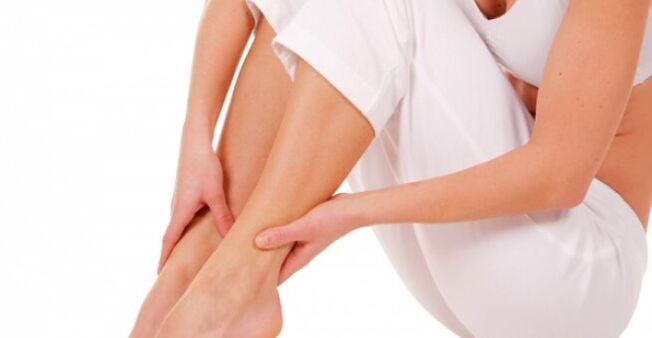
The first signs of the disorder are nonspecific (they are also present in other disorders) and they are grouped together under the term "heavy legs syndrome". It is characterized by increased and progressive lower extremity fatigue, leg pain, feeling heavy, calf burning and popping, and nighttime cramping of the calf muscles. These symptoms appear at the end of the day, especially if a person is standing or sitting for a long time during this time. Subsequently, as the pathology developed, nocturnal swelling of the back of the foot and ankle was added to the described disease manifestations. Leg soreness usually improves with rest.
Visual changes in the early stages of the disease are not always obvious because leg varicose veins start in deeper blood vessels. The only external sign of a problem that has started may be the network of blood vessels. Of course, they don't always indicate varicose veins, but when they do, it's best to consult a phlebologist, a venous disease specialist.
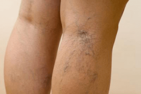
But in the later stages of varicose veins, cyanotic subcutaneous veins and varicose lymph nodes already appear - these are enlarged and tortuous superficial veins that resemble grapes. They are usually located on the calves and inner thighs.
Also, as the pathology progresses, the legs begin to swell more. A chronic venous insufficiency gradually develops, in which venous outflow and microcirculation in the tissues are disturbed. All of this is reflected in the condition of the skin on the legs: it darkens, peels, itches, and then a trophic ulcer develops, which heals poorly. This is how varicose veins develop. Similar varicose vein outcomes can be prevented with prompt treatment, so even mild but systemic discomfort in the legs, vascular network or "stars" on the skin, you should consult your doctor.
Symptoms of pelvic varicose veins
In the pelvis, varicose veins are less common than in the legs and occur mostly in younger women. The trigger for the development of this pathology is pregnancy (where both hormonal and mechanical factors come into play). After childbirth, signs of illness usually disappear, and only about 10% of women notice a periodic return of unpleasant symptoms after prolonged standing, hypothermia, and physical exertion.
Small pelvic varices manifest as chronic pelvic pain and dilation of the superficial veins of the perineum and vulva. Such patients are often unsuccessful in treating inflammatory diseases of the reproductive organs because lower abdominal pain (a characteristic of pelvic varices) is sometimes erroneously associated with chronic oophoritis, salpingitis, endometriosis, etc.
How are varicose veins diagnosed?
When varicose veins become clearly visible on a patient's legs, doctors can diagnose "varicose vein disease" even without the results of an instrumental study. In-depth examination is essential if the lesion has just begun to develop or is confined to the small pelvis.
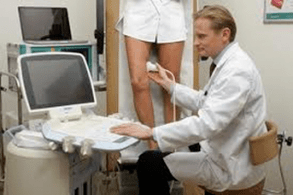
The primary method of diagnosing varicose veins is Doppler ultrasonography. This study provides information on venous lesions anywhere in the body. With the help of ultrasound, doctors can study the condition of the walls and the anatomy of the deep and superficial veins, valves, assess the blood flow in the blood vessels, detect the return of blood, etc. The classification of varicose veins and the selection of treatment methods are based on ultrasound findings.
Another diagnostic method used in this pathology is rheology. Its implementation allows you to determine the extent to which the tissues of the lower extremity are filled with blood and nutrients. This information helps the doctor determine what stage the disease is in: in the compensation stage, sub-compensation stage, etc.
Rarely, phlebography is used for varicose veins - this is an X-ray of the veins using a contrast agent.
In addition, a thorough examination of a patient with varicose veins usually includes various blood tests: the doctor is particularly interested in the levels of hemoglobin, red blood cells, platelets, and clotting parameters. These data make it possible to assess blood density and the tendency of a patient's body to form clots.